All published articles of this journal are available on ScienceDirect.

3D Printing in Maxillofacial Tissue Regeneration: A Review
 Mahsa Karkhaneh2
Farzin Ahmadpour1
Mahsa Karkhaneh2
Farzin Ahmadpour1
Authors Info & Affiliations
Abstract
Introduction
Three-dimensional bioprinting has changed tissue engineering in dentistry. It aids in creating precise tissue structures. This review focused on the developments in three- bioprinting for dental and maxillofacial tissue engineering.
Methods
This review examined previously published studies on bioprinting techniques used in dental and maxillofacial tissue engineering. Particular attention was given to the composition of bioinks, the types of cells employed, and the printing strategies used to fabricate tissue constructs. In addition, the available studies were evaluated to identify successful outcomes, existing limitations, and the major challenges associated with translating three-dimensional bioprinting into clinical practice.
Results
The findings highlight several commonly used bioinks, including gelatin methacrylate (GelMA), alginate, and collagen. These biomaterials are often combined with stem cells such as dental pulp stem cells and mesenchymal stem cells to support tissue regeneration. Recent advances in bioprinting techniques, particularly extrusion-based and inkjet printing, have enabled the fabrication of constructs with potential applications in pulp–dentin regeneration, alveolar bone repair, and oral mucosal reconstruction. Three-dimensional bioprinting allows the generation of highly precise tissue structures that more closely mimic the architecture of native tissues.
Discussion
Three-dimensional bioprinting shows considerable promise for future therapeutic applications in dentistry and maxillofacial reconstruction. This technology provides the ability to fabricate tissue constructs that replicate the structural and biological characteristics of native tissues
Conclusion
Continued research is required to optimize bioink formulations, improve printing accuracy, and facilitate the translation of three-dimensional bioprinting technologies into clinical practice for dental and maxillofacial tissue regeneration.
1. INTRODUCTION
Regeneration of oral and dental tissues is an important challenge in treating problems such as injuries, infections, congenital abnormalities, and age-related degeneration. Common treatment methods such as tissue grafting, dental implants, and root canal therapy usually cannot fully and naturally restore the tissue, and they also have limitations in long-term function and biological compatibility [1, 2]. Regenerative dentistry aims to repair and restore the structure and function of dental tissues that have been lost due to disease, trauma, or congenital defects. Traditional methods are often not successful in completely rebuilding the complex structures of teeth and may involve problems such as damage at the graft donor site, limited availability of suitable tissue, and the risk of immune rejection [3, 4].
Three-dimensional bioprinting has emerged as a promising approach in dental tissue engineering due to its ability to precisely control cell distribution and scaffold architecture. This spatial accuracy makes it particularly well suited for regenerating complex dental tissues [5, 6].Over recent years, substantial progress has been achieved in the development of advanced bioinks, specialized bioprinting platforms, and optimized cell sources tailored for dental applications. Interdisciplinary collaboration among materials scientists, stem cell biologists, and clinical dentists has significantly accelerated preclinical research and technological refinement [7, 8]. These advances enhance the suitability of three-dimensional bioprinting for addressing the heterogeneous microenvironment of the oral cavity. Despite these encouraging developments, several biological, technical, and regulatory challenges continue to limit the routine clinical implementation of three-dimensional bioprinting in dentistry.
A major factor driving interest in this field is the ongoing shift toward precision and personalized medicine. With the widespread integration of advanced imaging modalities, CAD/CAM systems, and three-dimensional digital modeling in routine dental practice, the clinical adoption of bioprinting technologies may become increasingly feasible. Such integration would enable the fabrication of patient-specific tissue constructs tailored to individual anatomical and biological characteristics. Ultimately, this personalized approach has the potential to improve regenerative outcomes and enhance overall treatment success [9, 10]. Three-dimensional bioprinting has the potential to give us treatments in dentistry and maxillofacial reconstruction.
Over the past decade, researchers have increasingly explored the use of three-dimensional bioprinting for the regeneration of various dental tissues. These include the pulp–dentin complex, periodontal ligament, alveolar bone, and oral mucosa. Bioprinting provides precise control over cell placement and scaffold architecture, making it a promising strategy for dental tissue engineering. Despite these advances, several challenges remain. These include the development of suitable bioinks, the promotion of vascularization within printed constructs, and ensuring effective integration with surrounding host tissues. In addition, issues related to safety, efficacy, and regulatory approval must be carefully addressed before clinical application can be achieved. In the context of trauma reconstruction, further considerations include the scalability of bioprinting processes and the mechanical strength required for functional tissue repair [11, 12].
Despite these advances, several challenges persist, including the development of optimized bioinks, the establishment of adequate vascularization within engineered tissues, and the functional integration of printed constructs with native host tissues. Furthermore, the translation of bioprinting strategies into clinical practice requires rigorous evaluation of safety, therapeutic efficacy, and compliance with regulatory frameworks. In the context of maxillofacial trauma reconstruction, additional considerations involve the need for time-efficient fabrication processes and the mechanical stability and load-bearing capacity of the fabricated scaffolds [ 13 ].
Recent studies have demonstrated significant progress in the application of three-dimensional bioprinting for tissue regeneration. Dixit et al. [ 14 ] reported that three-dimensional bioprinting enables the fabrication of patient-specific scaffolds with precisely controlled architectures and spatially organized cell distributions. Such capabilities are particularly beneficial for reconstructive procedures following trauma or tumor resection. Their findings also highlight the crucial role of biomaterials and cell-laden bioinks in promoting bone regeneration and facilitating tissue integration [ 14 ]. Similarly, Marini et al . emphasized the importance of integrating advanced imaging technologies with digital workflows for customized scaffold design and surgical planning. Their study demonstrated that imaging-guided bioprinting can enhance the accuracy of defect reconstruction and support the development of regenerative strategies for maxillofacial tissues [ 15 ].
This review aims to provide an overview of the current status of three-dimensional bioprinting in dental and maxillofacial tissue regeneration. It highlights key studies, discusses existing challenges, and outlines future directions in this rapidly evolving field. Figure 1 illustrates the potential application of three-dimensional bioprinting in maxillofacial trauma repair.
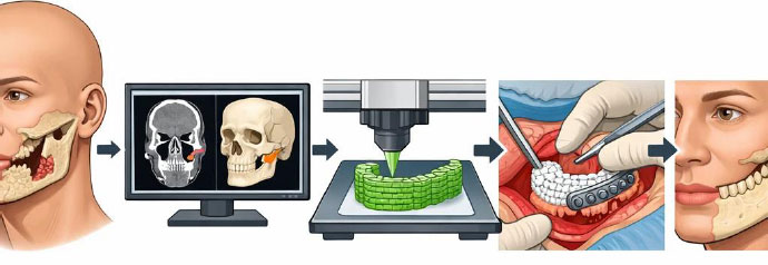
The use of three-dimensional bioprinting for maxillofacial trauma reconstruction.
1.1. Search Strategy and Databases
A comprehensive literature search was conducted to identify studies investigating the application of 3D printing in maxillofacial tissue regeneration. The electronic databases PubMed/MEDLINE, Scopus, Web of Science, and Google Scholar were systematically searched for relevant publications up to January 2026.
The search strategy included the following keywords and their combinations: 3D bioprinting, maxillofacial trauma reconstruction, dental tissue engineering, bioink, regenerative dentistry, maxillofacial regeneration, alveolar bone, and clinical translation.
Only articles published in English were considered eligible for inclusion. In addition, the reference lists of all selected studies were manually screened to ensure that no relevant publications were overlooked. This approach aimed to provide comprehensive coverage of the available literature on 3D printing in maxillofacial tissue regeneration.
1.2. Principles and Techniques of 3D Bioprinting
3D bioprinting is based on additive manufacturing. In this method, biomaterials and cells are placed layer by layer to create a tissue structure. There are several main bioprinting methods, including inkjet printing, extrusion printing, and laser-assisted printing. Inkjet bioprinting is fast, but it only works with low-viscosity bioinks. Extrusion bioprinting can use different materials and a higher number of cells. Laser-assisted bioprinting has very high accuracy and is suitable for making detailed dental structures, but it is expensive and more complex to use [16, 17].
One of the most important parts of bioprinting is the bioink. A bioink should be compatible with the human body. It should also help cells survive and grow. In addition, it needs suitable mechanical strength and should gradually degrade in the body. In dentistry, hydrogels such as alginate, gelatin methacrylate (GelMA), collagen, and decellularized extracellular matrix are commonly used [18, 19]. These materials can act similarly to the natural environment of dental tissues.
Living cells are incorporated during the bioprinting process to promote tissue formation. In most cases, these cells are stem cells with the capacity to differentiate into dental tissues [ 20 ]. Commonly used cell types include dental pulp stem cells (DPSCs), mesenchymal stem cells (MSCs), and induced pluripotent stem cells (iPSCs). In addition, growth factors such as bone morphogenetic protein-2 (BMP-2), vascular endothelial growth factor (VEGF), and transforming growth factor-β (TGF-β) are incorporated into the bioink or applied after printing to enhance tissue regeneration and repair. A major advantage of bioprinting is the precise spatial control over the distribution of cells and biological components, which enables the fabrication of complex and functional tissue constructs [ 21 ].
1.3. 3D Bioprinting in Dental Tissue Regeneration
3D bioprinting is used in dentistry for the regeneration of hard and soft tissues [22, 23]. One of its most important applications is the regeneration of the dentin–pulp complex. In this method, dental pulp stem cells (DPSCs) are used. These cells are placed inside a bioink that contains materials that support blood vessel formation and dentin formation. Studies have shown that this method can create tissue similar to dental pulp. This tissue contains blood vessels and can help keep the pulp alive. In the future, this method may help treat necrotic pulp and prevent tooth loss [24]. 3D bioprinting is also used for alveolar bone regeneration. In this method, hydrogels or scaffolds containing bioceramic materials and bone-forming cells are used. These structures can help form new bone [25]. Compared with traditional bone grafts, these structures are more compatible with the body. Their degradation rate can also be controlled, and they can be designed to match the shape of bone defects. This is important in injuries and periodontal diseases.
Regeneration of oral soft tissues, such as the gingiva and oral mucosa, is another application of this technology [26]. Bioprinting can create tissues that are used to cover oral wounds or large oral defects. These tissues can also be used in laboratories to study diseases such as oral ulcers and oral cancer. Most of these studies are still in the early stages, but in the future, they may replace or support tissue grafts [27]. Bioprinting can also produce multilayer tissues similar to natural oral mucosa. In this method, keratinocytes and fibroblasts are printed using hydrogels such as collagen and fibrin. These tissues have proper layering and can act as a protective barrier. For this reason, they are useful for oral mucosa regeneration and disease studies [28].
1.4. Maxillofacial Trauma Reconstruction
Recent efforts have focused on using 3D bioprinting to address trauma-induced defects in the maxillofacial region, including complex mandibular fractures and bone loss following tumor resection or accidents. Customized bioprinted scaffolds embedded with osteoprogenitor cells have been proposed to fit irregular bony geometries and promote vascularized bone regeneration. These constructs show potential in replacing traditional titanium plates or autografts, offering better integration and less donor-site morbidity [29].
1.4.1. Preclinical Studies
Yu et al. [30] explored the influence of alginate/gelatin (Alg-Gel) hydrogel scaffolds on the proliferation and differentiation of human dental pulp stem cells (hDPSCs). Their study convincingly demonstrated that 3D-printed Alg-Gel scaffolds provide a superior microenvironment for hDPSC growth and adhesion compared to traditional Alg-Gel scaffolds. This improvement was linked to the increased calcium and phosphorus ion content in the 3D-printed scaffold extracts, which in turn enhanced osteogenic and odontoblastic differentiation markers. While this work underlines the potential of 3D bioprinted hydrogels in supporting dental pulp regeneration, it is important to note that the study primarily relied on in vitro assays. The extrapolation of these findings to in vivo conditions requires further validation, especially regarding long-term scaffold stability and integration within the complex oral milieu. Additionally, the impact of scaffold degradation dynamics and mechanical properties on cell differentiation was only superficially addressed, signaling a need for more comprehensive biomechanical assessments [30]. This method can be adapted for pulp regeneration strategies in teeth affected by trauma or infection.
Han et al. [24] advanced the field by successfully fabricating a patient-specific 3D dentin-pulp complex through localized differentiation of hDPSCs using fibrin-based bioinks co-printed with polycaprolactone (PCL). This approach uniquely combined soft and hard tissue elements within a single construct, achieving spatially controlled odontogenic differentiation. The study’s emphasis on tuning fibrinogen concentration to optimize printability and cell viability highlights an important technical consideration often overlooked in bioprinting protocols. However, despite the promising localized mineralization after 15 days of culture, the study did not extensively evaluate the mechanical resilience of the construct under physiological loading conditions, which is crucial for dental applications. Moreover, the absence of in vivo testing limits our understanding of the construct’s immunogenicity, vascularization potential, and long-term functionality. The reliance on a differentiation medium also raises questions about how effectively the bioink itself can promote regeneration without exogenous factors, a key challenge for clinical translation [24]. This approach could be further adapted to regenerate dentoalveolar tissues damaged due to traumatic injuries or surgical resections.
Iranmanesh et al. [31] introduced an innovative alginate-gelatin hydrogel scaffold fabricated using UV-curable bioinks and coated with naproxen to modulate mechanical and biological properties. Their findings revealed that naproxen coating enhanced tensile strength and polymer adhesion, while maintaining biocompatibility and avoiding an acidic microenvironment during degradation. This study is notable for integrating drug delivery within the bioprinted scaffold, addressing a critical need for multifunctional constructs capable of both tissue regeneration and inflammation control. Nonetheless, the work primarily focused on mechanical and physicochemical characterizations, with limited insight into cellular responses specific to dental tissues. The increase in degradation rate associated with naproxen release also warrants careful balancing to ensure scaffold longevity aligns with tissue healing timelines. Importantly, while the UV-curing process offers rapid scaffold fabrication, its effects on encapsulated cell viability and differentiation potential were not investigated, highlighting an essential gap in assessing the full regenerative capacity of the material [31]. Their scaffold design may also have applications in craniofacial bone reconstruction where inflammation modulation is critical post-trauma.
The strong odontogenic and angiogenic potential of human dental pulp stem cells has long been established, with early seminal studies demonstrating their capacity to regenerate pulp–dentin–like tissues in vivo [32]. Recent insights have highlighted the central role of biomaterial composition and microenvironmental cues in directing hDPSC fate, particularly in the context of dentin–pulp tissue engineering [33].
A randomized controlled trial by Nicot et al. [34] investigated the educational impact of 3D-printed anatomical models in teaching craniofacial trauma. The study highlighted the limitations of traditional two-dimensional visuals in conveying the spatial and biomechanical complexities of facial injuries. By comparing medical students’ comprehension after using either printed 3D models or standard 2D images, the research demonstrated a significant advantage of 3D tools in improving both morphological understanding and biomechanical reasoning. Notably, students engaging with 3D models performed better on assessments involving complex fractures such as zygomatic and mandibular breaks. Although not directly focused on bioprinting for reconstruction, this evidence underscores the broader utility of three-dimensional fabrication technologies in enhancing anatomical literacy-an essential prerequisite for advancing regenerative strategies in maxillofacial trauma care [34].
In a clinical series conducted by Ghantous et al. [35], additive manufacturing (AM) was employed in the surgical management of 16 maxillofacial patients over five years, with nearly half of the interventions addressing trauma-related or post-traumatic conditions. The study demonstrated the practical versatility of 3D-printed, patient-specific implants (PSIs)-particularly in orbital floor reconstructions-highlighting titanium as the predominant material. While the technology facilitated accurate preoperative planning and personalized reconstruction, challenges such as intraoperative misfit and minor postoperative complications, including edema and implant exposure, were reported. Notably, PEEK-based reconstructions were utilized for cases involving more complex bone regions like the nasal, temporal, and frontal areas. Despite some limitations, the findings underline the clinical relevance of 3D printing in trauma care, especially in producing custom implants that conform to individual anatomical defects. These insights offer a valuable precedent for extending the scope of 3D bioprinting from structural modeling to biologically integrated reconstruction in facial trauma [35].
1.4.2. Review Studies
Oley et al. [36] conducted a systematic review evaluating the role of three-dimensional printing in craniomaxillofacial (CMF) trauma reconstruction, analyzing 20 human studies involving 170 patients. The review underscores the growing reliance on 3D-printed solutions-ranging from anatomical models to patient-specific implants and customized surgical tools-as alternatives to conventional graft-based methods. The outcomes across most studies were favorable, reporting improvements in both aesthetics and function, such as restoration of facial symmetry, orbital volume correction, and enhanced implant fitting. Importantly, the review notes a reduction in intraoperative time due to improved pre-surgical planning, though offset by longer preparation times and increased manufacturing costs. While the technology shows clear promise, particularly in improving surgical precision and patient outcomes, barriers remain-especially in low-resource settings where access to advanced printing capabilities is limited. This synthesis reinforces the potential of 3D printing in CMF trauma care and sets the stage for integrating bioprinting as a future step toward regenerative, patient-specific solutions [36].
In their review study, Park et al [37] reported that the mandible plays a critical role as a dynamic bio-organ, providing structural support to the lower face while facilitating essential functions such as mastication and speech [37]. Extensive mandibular defects-typically those exceeding 6 cm-often necessitate complex reconstructive strategies like osteocutaneous vascularized free flaps. While effective, these techniques are constrained by donor site morbidity and the inherent burden of additional surgical procedures. In response, recent advancements in three-dimensional (3D) bioprinting have emerged as a promising alternative, aiming to replicate both the anatomical and functional characteristics of composite mandibular tissues. Key elements in 3D bioprinting for bone reconstruction include scaffolds, viable cells, and bioactive factors. For mandibular applications specifically, the printed constructs must offer sufficient mechanical integrity, structural resilience, and biocompatibility to withstand functional loads. Recent innovations have introduced multi-cartridge bioprinting platforms capable of simultaneously depositing cell-laden hydrogels and structural polymers, thus reinforcing mechanical performance while promoting tissue integration. To achieve clinically viable outcomes in mandibular bioprinting, future approaches should integrate biological requirements with digitally driven customization platforms. Such systems would support personalized, on-demand fabrication of tissue constructs, offering a scalable and potentially more regenerative solution to large mandibular defects [37].
Table 1 summarizes key studies on 3D bioprinting in craniofacial trauma and mandibular reconstruction
| Study Type | Authors (Year) | Focus/Intervention | Key Findings | Limitations/Notes |
|---|---|---|---|---|
| Preclinical | Yu et al. [30] | Alginate–gelatin scaffold with hDPSCs | Enhanced cell proliferation, osteogenic/odontogenic differentiation in vitro | No in vivo validation; limited mechanical analysis |
| Han et al. [24] | Fibrin-based bioink + PCL for dentin–pulp regeneration | Spatially controlled differentiation; good viability and mineralization | No in vivo data; mechanical testing under loading conditions is absent | |
| Iranmanesh et al. [31] | Naproxen-coated Alg-Gel scaffold via UV-curing | Improved mechanical strength and anti-inflammatory potential | Cellular response and UV impact on viability were not assessed | |
| Nicot et al. [34] | 3D-printed models for trauma education | Improved understanding of facial trauma anatomy and biomechanics in students | Not directly on reconstruction; educational focus | |
| Ghantous et al. [35] | Clinical use of 3D-printed PSIs in maxillofacial trauma | Effective reconstruction; titanium and PEEK implants; some complications | Small sample; technical complications noted | |
| Review | Oley et al. [36]. | Systematic review on 3D printing in CMF trauma | Favorable outcomes in esthetics/function; reduced surgical time | Cost and access barriers in low-resource settings |
| Park et al. [37] | Review of mandibular bioprinting strategies | Emphasized the need for strong, biocompatible scaffolds and customizable platforms | Conceptual; lacks experimental/clinical data |
1.5. Challenges
Despite the advances made in 3D bioprinting, certain hurdles still need to be overcome before the technology can be translated into clinical use. The design and synthesis of appropriate bioinks that can emulate the native extracellular matrix while maintaining cell viability, proliferation, and differentiation is one such challenge. Vascularisation of bioprinted constructs is also a significant issue [38].
In cases of trauma, the time-dependent reconstruction and mechanical load-carrying function of bioprinted scaffolds become an additional concern, demanding a very fast timescale of fabrication and high structural stability [39]. Aspects of other concerns of printed constructs, like integration with the host tissues, immune compatibility, and functionality over long periods of time, have to be taken into consideration.
Apart from cost considerations, there are implications of regulation and ethics that hinder the translation of bioprinted products into the clinic. There is a requirement to generate guidelines for standardized bioink compositions, printing conditions, and maturation conditions [40].
On future directions, research should be directed at the bioprinting of multifunctional bioinks, improving techniques for bioprinting of vascular networks, and conducting in vivo, preclinical, and clinical studies testing bioprinted constructs for both safety and efficacy. The future success of 3D bioprinting in dental and craniofacial tissue engineering will depend on the collaborative efforts between material scientists, biologists, engineers, and clinicians.
1.6. Comparative Overview of Bioinks used in Maxillofacial Bioprinting
Various natural and synthetic materials have been utilized as bioinks in maxillofacial and dental tissue bioprinting, each with distinct physicochemical and biological properties. Natural polymers such as collagen, gelatin methacrylate (GelMA), alginate, and fibrin provide excellent biocompatibility and support cell adhesion and proliferation [41]. For example, collagen and GelMA hydrogels promote odontogenic differentiation of dental pulp stem cells due to their similarity to native extracellular matrix composition. However, these soft hydrogels often exhibit limited mechanical strength and rapid degradation rates, restricting their application in load-bearing tissues like alveolar bone [41].
Synthetic polymers, including polycaprolactone (PCL), polyethylene glycol diacrylate (PEGDA), and bioactive ceramics such as hydroxyapatite or β-tricalcium phosphate (β-TCP), improve structural stability and enable precise architectural control [42]. These materials demonstrate good printability and form retention but may require surface modification or blending with natural polymers to enhance cellular viability and biological integration.
Hybrid bioinks-combinations of natural and synthetic components-offer a balance between biological performance and mechanical stability [43]. For instance, Alginate–GelMA composites maintain high cell viability (>90%) up to 7 days post-printing while improving elastic modulus compared to pure gelatin systems. Similarly, the incorporation of nano-hydroxyapatite into GelMA or PCL matrices promotes osteogenic differentiation and mineral deposition, making them ideal for bone regeneration [43].
Table 2 shows a summarized comparison of frequently used bioinks and their key properties [41-43].
| Material | Type | Cell Viability | Proliferation Support | Mechanical Strength | Degradability | Typical Application |
|---|---|---|---|---|---|---|
| Collagen | Natural | Excellent | High | Low | Fast | Soft tissue, pulp regeneration |
| GelMA | Semi-synthetic | Excellent | High | Moderate | Controlled | Dentin–pulp, mucosal |
| Alginate | Natural | Good | Moderate | Low | Variable | Scaffold matrix, hybrid bioink |
| Fibrin | Natural | Excellent | High | Low | Fast | Pulp, soft tissue repair |
| PCL | Synthetic | Moderate | Low | High | Slow | Bone, structural support |
| PEGDA | Synthetic | Moderate | Moderate | High | Controlled | Bone, cartilage models |
| Hydroxyapatite / β-TCP | Ceramic | N/A (acellular) | N/A | Very High | Slow | Alveolar bone reconstruction |
1.7. Clinical Translation and Regulatory Considerations
To make the leap from laboratory discoveries to clinical application, a coordinated effort by researchers, the industry, and regulatory agencies will be necessary. An important route to this end is the utilization of bioprinted constructs to facilitate personalized surgical planning and use during intraoperative guidance [44]. Bioprinted models of dental defects and jawbone can allow better preoperative planning and prediction of outcomes for reconstructive surgery [45].
New translational research is now beginning to investigate pilot clinical use of bioprinting techniques, Most of all in the field of reconstructive and maxillofacial surgery. Despite this, there are currently limited clinical trials, with most study types being confined to either case studies or animal models [46].
Factors on immunogenicity, biodegradation, and ability to withstand functional load long-term will need investigation before any large-scale human trials [46]. Bioprinted products are also difficult to regulate. Being either a device, biologic, or combined product, the bioprinted sample can have a different pathway to approval. Although the FDA and EMA are formulating policies, the standards and procedures need to be finalized. Collaboration between regulators, academia, and industry is needed to develop a way to deploy them safely in clinics [47].
1.8. Future Perspectives and Conclusions
The future is promising for 3D bioprinting applications for dental tissue regeneration and repair if various interdisciplinary challenges can be overcome. 3D printing techniques combined with the rapid progression of biomaterial design, in particular smart and sensitive hydrogels, will enable the production of dynamic constructs to meet the demands of the constantly changing oral environment. Also, bioprinting techniques combined with online image-guided surgical planning and computer-generated AI design algorithms are expected to increase therapeutic success.
1.9. AI in Maxillofacial Reconstruction and Regeneration
The application of artificial intelligence (AI) in maxillofacial reconstruction and regeneration may provide a new approach to optimize surgical planning, enhance the accuracy of bioprinting, and enable a personalized treatment strategy. AI algorithms analyze large amounts of data to find the best biomaterials and design constructs that conform to an individual patient's anatomy. Like that, machine learning models can be used to estimate the predicted outcome as preoperative imaging and patient factors, which offer promise to prevent complications and maximize the success rate. On top of that, AI-enabled tools may be used to monitor and evaluate the progress of tissue regeneration dynamically to derive a more adaptive strategy. As AI technology progresses, the use of AI in the clinical aspect of maxillofacial reconstruction and regeneration might be able to break down the barriers of bioprinting technology and personalized medicine [48]. Figure 2 shows AI in maxillofacial reconstruction and regeneration.

AI in maxillofacial reconstruction and regeneration.
New technologies such as four-dimensional (4D) bioprinting have created new opportunities in regenerative dentistry [47]. In this method, the structure or function of the printed tissue can change over time. This feature may be useful for developing advanced implants and better tissue repair methods. In addition, using different types of cells together with blood vessels and neural elements can make the printed structures more similar to natural body tissues.
3D bioprinting has a promising future in regenerative dentistry and maxillofacial reconstruction. This technology allows the precise fabrication of tissues with desired shapes and cellular compositions. Because of this, tissues can be repaired in a more accurate and patient-specific way. However, more research and technological development are still needed before this method can be widely used in clinical treatment. With further progress, bioprinting may become one of the main approaches in regenerative dentistry and maxillofacial reconstruction and help repair complex tissue defects.
LIMITATION
3D bioprinting's advances have been quite rapid in the last few years, but several issues still need to be overcome before it can be a routinely used treatment. The main obstacle is the inability of printed tissues to generate an adequate vascular and neural network. This is important because cells need a continuous supply of nutrients and oxygen, and if an inadequate blood supply is generated, the tissue may not survive very long once implanted.
The printing materials also pose issues. Soft bioinks lack the resilience required in an area that may be subject to movement or pressure.
Some materials may degrade too quickly or require longer than expected to be in the body, both impacting levels of healing and tissue attachment. A second problem is that the results are not always reproducible. Sometimes a tissue will perform very efficiently in one experiment but not in a second.
Producing huge quantities of tissues with uniform quality remains expensive and continues to be a problem. Approval by a clinical regulatory body remains another hitch since all new materials will have to go through detailed safety tests before they can be introduced to patients.
AUTHORS’ CONTRIBUTIONS
All authors contributed to data collection, the drafting, and scientific revision of the manuscript. All authors read and approved the final manuscript.